

Panoramic dental X-ray (OPG): uses, procedure and radiation dose
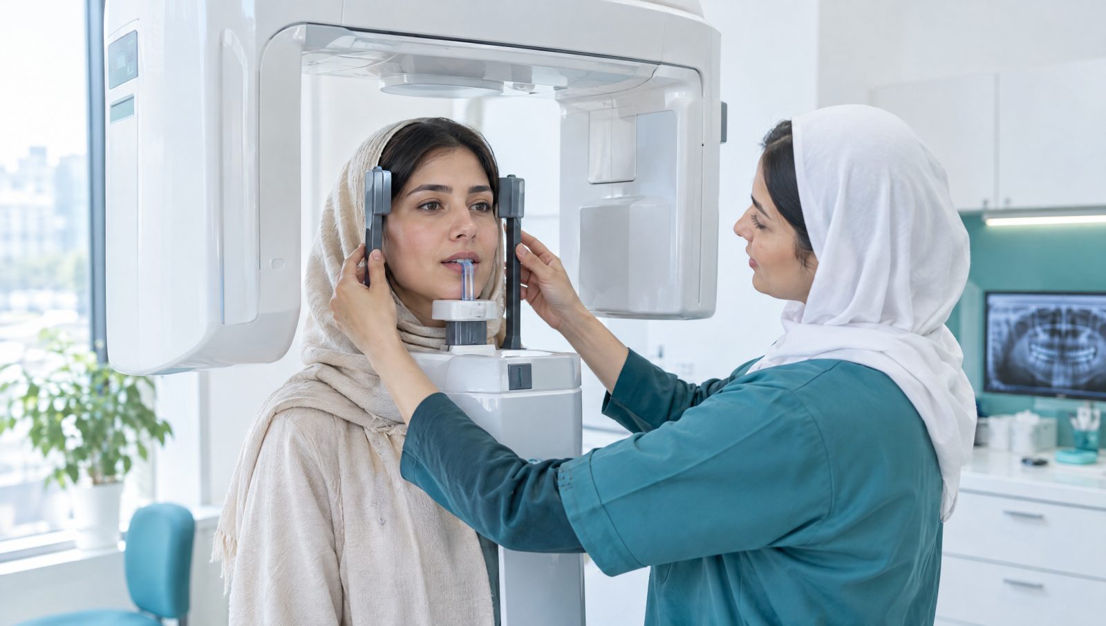
A panoramic dental X-ray shows every tooth and both jaws in a single image in a few seconds. Learn when it is needed, how it is taken, how much radiation it involves and how it differs from CBCT.
A panoramic dental X-ray, also called an OPG (orthopantomogram), is one of the most widely used images in dentistry. In a few seconds it shows every tooth, both jaws and much of the surrounding anatomy in a single picture, which is why many dentists use it as the first step in a general assessment. This guide explains what a panoramic X-ray is, when it is needed, how it is taken, how much radiation it involves and how it differs from CBCT.
What is a panoramic X-ray?
In panoramic radiography the X-ray tube and the image receptor rotate around your head at the same time, on opposite sides. The result is a wide, two-dimensional image that shows the upper and lower dental arches as if they had been unrolled. Unlike intraoral images, which show one or a few teeth, a panoramic image gives an overview of:
- all erupted and impacted teeth, including wisdom teeth
- the bone of the upper and lower jaws
- both temporomandibular joints (TMJ), in general terms
- the floor of the maxillary sinuses
- in many patients, the canal of the nerve in the lower jaw
When does a dentist request one?
- Wisdom teeth: to see how an impacted tooth is positioned and angled before deciding on extraction.
- Orthodontics: to check the number and position of teeth and roots before and during treatment, usually alongside cephalometric radiography.
- Children: to follow the development of permanent teeth and detect missing or extra teeth.
- Initial implant assessment: a first look at the bone; detailed planning usually needs CBCT.
- Gum disease: to see the overall pattern of bone loss around the teeth.
- Lesions and injuries: cysts and bony lesions, jaw fractures after trauma, and a first look at the jaw joints.
- Comprehensive examination: at a first visit or before extensive treatment, as a reference image.
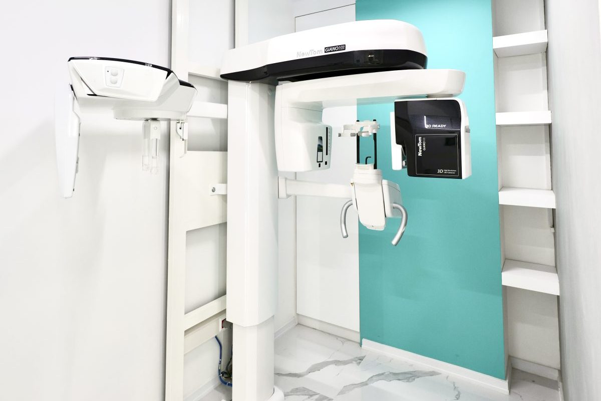
How the X-ray is taken
The whole visit usually takes only a few minutes, and the exposure itself lasts only while the unit rotates — roughly ten to twenty seconds.
- Remove metal items from your head and neck: earrings, necklaces, hair clips, glasses and removable dentures.
- Stand or sit in front of the unit and rest your chin on the support.
- Gently bite on a disposable plastic block so your jaws are in the right position.
- The technician steadies your head with the side guides.
- Close your lips and press your tongue against the roof of your mouth — this simple step prevents a dark shadow over the roots of the upper teeth.
- Stay completely still while the unit rotates. Even small movements can distort the image and mean a repeat, and therefore a second exposure.
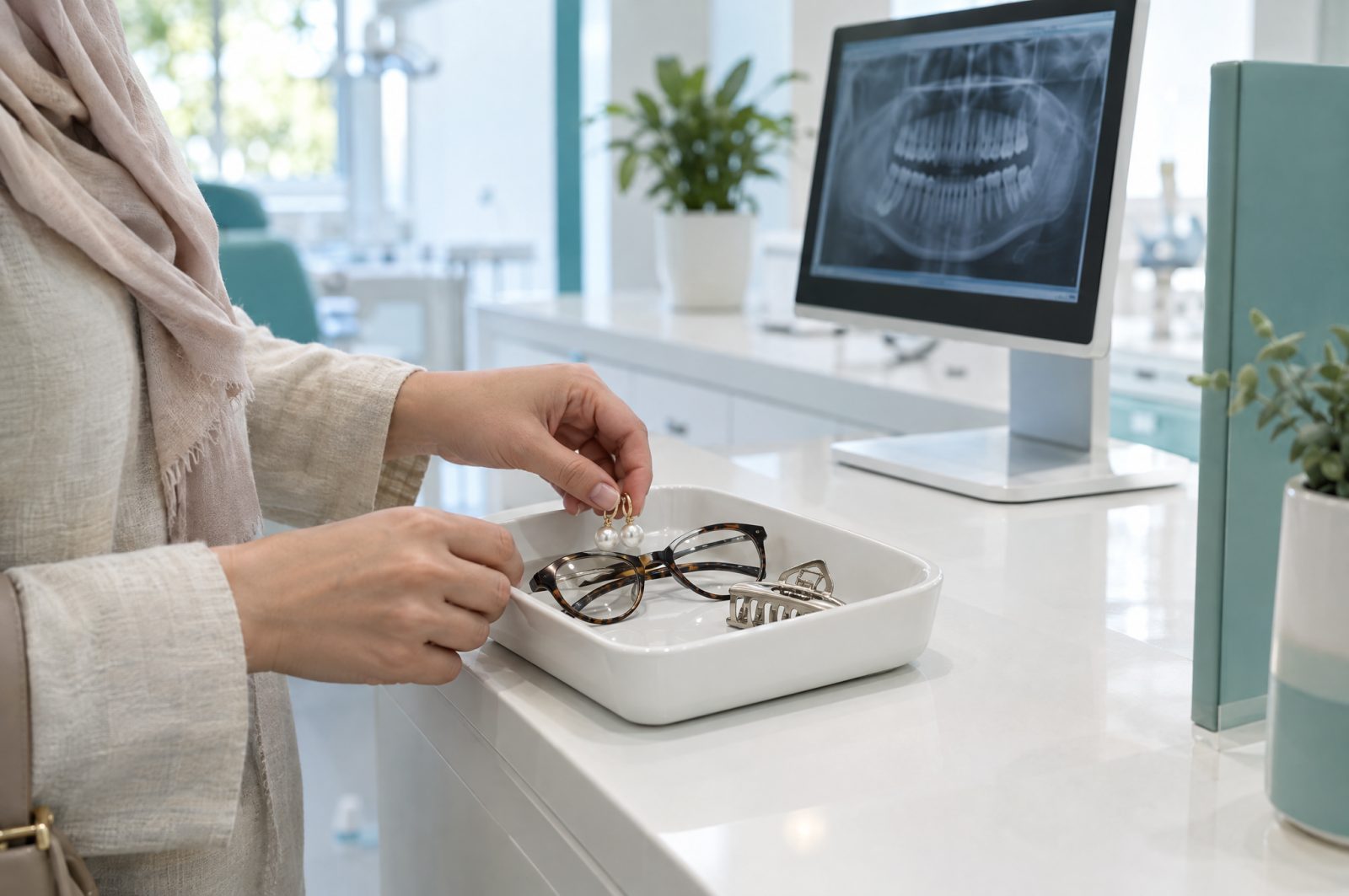
Preparing for your panoramic X-ray
No special preparation such as fasting is needed. A few simple steps make the visit quicker and the image better:
- Bring the written request from your dentist.
- Bring any previous images — they sometimes make a repeat unnecessary.
- If you are using insurance, bring the documents you need, and check our contracted insurers beforehand.
- If you are, or may be, pregnant, tell us before the scan.
For a fuller checklist, read how to prepare for your imaging appointment.
How much radiation is a panoramic X-ray?
The effective dose of a digital panoramic X-ray is typically around 16 microsieverts, depending on the unit and its settings. To put that in context:
- it is roughly two to three days of the natural background radiation we all receive;
- it is about the same as the cosmic radiation of a four-hour flight;
- it is less than a sixth of the dose of a simple chest X-ray.
A low dose is still not a reason to image without cause: under the ALARA principle every exposure must be clinically justified and kept as low as reasonably achievable. Compare your own imaging dose with the radiation calculator, or read how much radiation a dental scan involves.
Limitations of a panoramic X-ray
- It is two-dimensional: structures overlap, and the width of the bone from cheek to tongue cannot be seen.
- Magnification is uneven: it varies across the image, so it is not suitable for precise measurement.
- Less fine detail: small cavities between teeth and subtle changes at the root tip are better seen on intraoral periapical and bitewing images.
That is why a dentist may ask for an additional image after reviewing a panoramic. It does not mean the first image was inadequate — each method answers a different question.
Panoramic X-ray vs CBCT
| Feature | Panoramic (OPG) | CBCT |
|---|---|---|
| Image | 2D overview of both jaws | 3D, reviewable in three planes |
| Approximate dose | about 16 µSv | about 50–150 µSv, depending on field of view |
| Precise measurement | not suitable | possible and accurate |
| Main uses | overview, wisdom teeth, orthodontics, follow-up | implant planning, complex impactions, lesions, difficult root canals |
To learn more, read what CBCT is and how it differs from an ordinary dental X-ray.
Panoramic X-rays for children
Children have panoramic X-rays when there is a clinical reason, for example to check the eruption of permanent teeth or look for extra teeth. Exposure settings are adjusted to the child's size. The key requirement is that the child can stay still for a few seconds; explaining the steps simply and having a parent nearby usually helps.
Your results
The image is captured digitally and given to you or your dentist. When your dentist requests it, an oral and maxillofacial radiologist reviews the image and writes a specialist report that also covers findings outside the area of interest, such as sinus changes or silent jaw lesions. Referring dentists can download their patients' images and reports from the colleagues' portal.
Frequently asked questions
Do I need to fast before a panoramic X-ray?
No. You can eat and drink as usual before a panoramic X-ray.
How long does it take?
The exposure lasts about ten to twenty seconds while the unit rotates, and the whole visit usually takes only a few minutes.
Can I have a panoramic X-ray during pregnancy?
If it is clinically needed and cannot wait, it can be taken: the beam is aimed at the jaws and the dose reaching the uterus is negligible. Always tell us you are pregnant; the decision rests with your dentist. Read more in dental X-rays during pregnancy.
Does a panoramic X-ray show cavities?
Larger cavities, yes. Small cavities between teeth are seen much better on bitewing images.
Can I have one with implants or metal fillings?
Yes. Fixed restorations stay in place; only removable items such as dentures and jewellery need to come out.
To book a panoramic X-ray or any other oral and maxillofacial imaging, use the appointment form and our team will call you back.